62. DAH Symposium 2023
- 04.-06. Mai 2023
- Bad Homburg, Germany
- www.dah.at
PHOENIX 3000 replaces a conventional hand surgery table, while simultaneously boosting its functionality by an integrated X-ray-based imaging system.
A support arm above the operating field accommodates the X-ray source. The corresponding image receptor is built into the tabletop, on which the patient’s arm rests during the intervention. The motor-driven generator/detector assembly can be aligned exactly with the imaging object with the help of a laser targeting device.

A separate mobile monitor stand with a generously dimensioned dual-image flatscreen enables real-time monitoring of the intervention as well as side-by-side display of two fluoroscopic or video images for the purpose of comparison.
An integrated copper pre-filter brings about skin dose reductions of up to 30%, while an especially optimized software algorithm ensures consistently brilliant image quality. This makes the PHOENIX 3000 one of the few extremity X-ray systems that are suitable for pediatric applications in compliance with the EN 60601-1-54 /203.7.1 standard.
+ Automatic dose rate control (ADRC)
+ Positioning of the imaging object far from the X-ray source forced by intelligent design
+ Dose reduction through built-in copper pre-filter
+ Consistently high image quality thanks to sophisticated software algorithm
+ Fulfills the requirements for pediatric imaging
The imaging/surgery table of the PHOENIX 3000 is installed once prior to the beginning of an intervention next to the patient positioning table and will stay in its place during the entire procedure.
Depending on the patient’s arm length and the task to be solved, the imaging/surgery table may be placed at the side of the patient positioning table either with its column end or its open end. HANDY WORKPLACE LIGHTING Two LED strips mounted at the bottom of the generator support arm greatly support the OR team during patient preparation. STERILE COVERS

Support side

Open side
The motorized vertical adjustment of the imaging/surgery table is controlled by easily accessible buttons on both sides of the tabletop, allowing surgeons to lift or lower the table quickly and effortlessly to the height that is most comfortable for them.


Individual setting options
Two LED strips mounted at the bottom of the generator support arm greatly support the OR team during patient preparation.
For the most part, the sterile surgical drapes that are already on stock in the hospital or clinic may be used. Optionally, a made-to-measure sterile cover for the support arm is available.
An intuitive user interface and cleverly placed controls allow operators to familiarize themselves quickly and easily with the system.
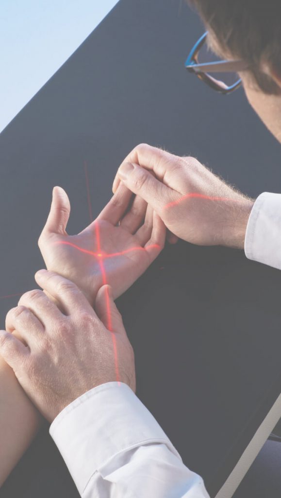
The field of view (FOV) is aligned precisely with the region of interest (ROI) with the help of a laser targeting device and without radiation. This is done by the surgeon only once prior to the intervention, thus eliminating the need for time-consuming repositioning involving additional radiation.
The multifunctional foot control keeps the surgeon’s hands free during an intervention, since all relevant operating functions – such as image and film sequence acquisition, folding up and down the generator support arm, saving or printing image data – can be controlled by means of foot pedals.

Practical multifunctional foot switch
A vertical free space twice as large as that of a conventional mini C-arm setup creates a highly convenient environment for surgery work. Fluoroscopy is possible even during drilling procedures, thus providing real-time visual feedback on the drill path. Folding back the generator support arm increases the OR team’s elbow room even more.

The acquired fluoroscopic images and film sequences are displayed enlarged on a high-resolution 27“ dual-image flatscreen. In addition to X-ray images and films, users may also permanently save external live views of the intervention (full-color single frames or entire movies), which are generated by the integrated video camera.
The video camera allows you to document surgical interventions from an external perspective. You can display color images in real time on the screen during an intervention and optionally save single frames or entire movies to the system’s hard disk.
The system features a cine loop function, thus enabling users to record and save fluoroscopic image sequences.
The system comes with a built-in USB interface for the connection of cordless USB storage devices. In this way you can export single images and film sequences in TIF or AVI format to external media and subsequently view or post-process them on any computer.
All generated still and moving images can be saved permanently to the system’s large internal hard disk. A clearly-organized patient and image database lets you retrieve all stored information rapidly and conveniently.
The system may be equipped with a video printer producing image printouts on thermal paper in 9 × 9 cm format.
The system supports the following DICOM classes:
• DICOM Worklist for importing patient data from a HIS / RIS
• DICOM Store for transferring and saving images to a PACS server
• Modality Performed Procedure Step (MPPS) for generating dose reports
+ Ample working space
+ Quality assurance and improvement thanks to different control views (e.g. sky view)
+ Relaxed surgery work and smooth workflow
+ Legally watertight documentation
+ Preservation of sterility, as it is no longer necessary to keep moving around a separate imaging unit
+ Perfectly matched, sturdy components
+ No repositioning necessary
+ Lower radiation burden
+ Shorter anesthesia times
+ Decreased risk of infections
+ All-in-one, compact and reliable X-ray system
+ No costly purchase of single pieces of equipment
+ Improved OR capacity utilization thanks to shorter intervention times
+ More space in the OR for other essential equipment
+ Less staff needed, since system can be handled by the surgery team alone
Distal Radius Fracture
Elevating the forearm results in the central ray beam penetrating the wrist at a 20–25° angle and hence parallel to the radiocarpal joint space.
Dorsopalmar View of the Wrist
Positioning of the forearm and the hand at shoulder height for dorsopalmar projections.
Lateral View of the Wrist
(Adduction of the upper arm and upright placement of the ulnar edge of the hand on the image receptor for lateral views of the wrist.
Palmar View of the Index
The dorsal side of the index finger rests on the image receptor. All other fingers are flexed.
Sky View
(1) In order to assess whether intra-articular screws are penetrating the radius in dorsal direction, the X-ray beam must be parallel to the longitudinal axis of the forearm.
(2) This is achieved by supinating the arm and flexing it to 90°, and then flexing the wrist.
In today’s ORs, crammed with all kinds of equipment and appliances, the two-in-one design of the PHOENIX 3000 saves valuable space. Its slim monitor stand will also easily find an appropriate place nearby. Despite its compact design, the imaging/surgery table provides an unprecedented movement range to hand surgeons, while its sturdy, perfectly balanced construction greatly facilitates precision surgery work. With the PHOENIX 3000, you are optimally prepared to meet any challenge.
In the emergency room, every second counts. The imaging/surgery table of the PHOENIX 3000 is instantly ready for use, avoiding you the hassle of arranging several different components and allowing you to deliver acute care to patients more quickly. With the PHOENIX 3000, no further equipment is required – all you need is included already!
In the treatment room, excellent image quality is of paramount importance for diagnostics and intervention planning. In addition to an advanced image processing system, details such as the built-in workplace lighting and laser targeting device assist you in obtaining accurate and information-rich scans. If necessary, you may record fluoroscopic cine loops and even video movies in order to visualize skeletal movements. With the PHOENIX 3000, you will instantly get a clear picture of the situation.
Allersberger Straße 185 / A7
90461 Nürnberg
Phone +49 911 4244967
Telefax +49 911 4244968
Mail info@medi-x.eu